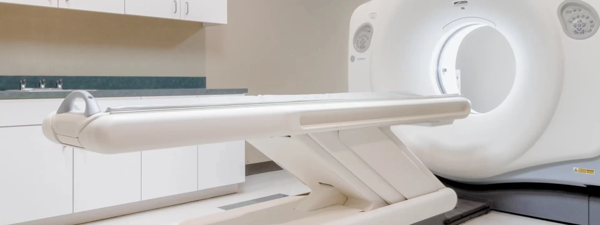
Nuclear Medicine Scan in Dallas, TX
At Crown Imaging, we provide Nuclear Medicine scans using a GE Nuclear Medicine Machine. Considered one of the leading technologies in this field, it is a highly advanced system designed to deliver precise and high-quality diagnostic imaging. These machines help doctors detect diseases at an early stage, improving patient outcomes.
What is Nuclear Medicine?
Nuclear Medicine is a diagnostic test in Radiology that uses certain pharmaceuticals that emit small doses of X-Rays and gamma rays that help physicians diagnose and treat disease. These radiopharmaceuticals are usually given either by way of an IV in the arm or by pill, where they are able to travel to specific organs that are being imaged with a camera able to detect the small doses of radiopharmaceuticals, generating digital images. These images are then given to a Radiologist to review.
Nuclear Medicine is unlike other Radiology tests such as CT, MRI, or Ultrasound in that Nuclear Medicine has the ability to identify both body function as well as the physical structure of certain organs instead of only identifying the physical structures of the organs that are imaged. This attribute gives the physician the advantage of detecting possible existing abnormalities at an earlier stage, before any physical changes of the organ have even occurred.
Unlike the case with Radiation Therapy – which uses radiation for treating disease – the amount of radiation that is contained in our test doses is minimal, much like an X-Ray procedure and therefore is safe.
Nuclear Medicine is a unique scan in the medical imaging community not available in most outpatient centers.
What is the Difference Between Nuclear Medicine Scan and X-Rays or CT Scans?
While X-Rays and CT scans capture anatomical images, nuclear medicine scans provide details information about organ function and metabolism.
How Does a Nuclear Medicine Scan Work?
Nuclear medicine imaging involves several key steps:
- Administration of Radiotracer: A small amount of radioactive material in introduced into the body via injection or oral ingestion.
- Absorption and Distribution: The radiotracer travels through the bloodstream to the targeted organ or tissue, accumulating in areas of interest.
- Conversion into Images: The GE Nuclear Medicine Machine detects gamma rays emitted by the radiotracer and converts them into detailed images.
- Image Processing and Diagnosis: A computer processes the data to create images that our on-site radiologist interprets to diagnose conditions.
Common Uses of Nuclear Medicine Scans
Nuclear medicine scans play a crucial role in diagnosing and managing a wide range of medical conditions including:
- Heart Imaging (Cardiology): Help evaluate blood flow to the heart muscle, helping detect coronary artery disease as well as assess heart function and ejection fraction for heart failure patients.
- Cancer Detection (Oncology): Identify cancerous tumors, determine their spread, often referred to as staging, and assess treatment effectiveness.
- Brain Imaging (Neurology): Used to detect Parkinson’s disease or identifies conditions such as Alzheimer’s disease and epilepsy.
- Thyroid Studies (Endocrinology): evaluate thyroid function, detect abnormalities such as hypothyroidism or nodules, and identify overactive parathyroid glands.
- Digestive System (Gastroenterology): Detects diseases such as bile duct obstructions and liver disease.
Benefits of Nuclear Medicine Scans
- Ealy Disease Detection: Can identify conditions before symptoms appear.
- Functional Imaging: Provides insight into organ function rather than just structure.
- Minimally Invasive: Requires only a small amount of radiotracer.
- Personalized Treatment: Helps doctors tailor treatments based on metabolic activity.
Safety Considerations
While nuclear medicine involves exposure to radiation, the amount is minimal and considered safe. The benefits of early diagnosis often outweigh the risks. Patients should follow any preparation instructions provided by their healthcare provider to ensure optimal imaging results.
Most common Nuclear Medicine studies:
There are three categories of studies:
Bone Scan:
A Nuclear Bone Scan is a diagnostic test that is conducted for the purpose of identifying certain abnormalities of bone such as fractures, infections, or tumors. During this test, you will be given an injection of a radiopharmaceutical that has no side effects into a vein in your arm or hand.
Depending on the instructions from your Physician, images of the affected area of your body that has bone pain/inflammation could be taken immediately after the injection. A whole-body scan will occur 3 hours after the injection. During the 3 hours wait time, you will be able to eat and drink plenty of fluids.
The entire procedure takes approximately 4 to 4 and 1/2 hours. It does not require you to fast or change into a hospital gown.
Physician Note:
Crown Imaging provides the extra service of including a whole-body scan in addition to the delay images of the limited area of interest
Hida Scan:
A Hepatobiliary Scan (Hida Scan) is a diagnostic test that is conducted for the purpose of evaluating gallbladder function. During this test, you will be given an injection of a radiopharmaceutical that has no side effects.
A Nuclear Medicine Technologist will take images as the radiopharmaceutical moves through your liver, gallbladder, and intestines. This usually takes an hour.
Once the radiopharmaceutical has reached the liver, gallbladder, and intestines, you will receive an additional injection of a medicine that simulates a fatty meal. This medicine is used to measure the function of your gallbladder.
The entire procedure typically takes 2 to 2 and ½ hours. In order to prepare for this test, you will need to fast from food and water for at least 6 hours before the test and it is also important to consult with your physician about any of your regular medications that you may or may not be permitted to take on the day of the test. After the test, you will be able to resume normal activity such as eating and drinking.
MUGA :
A Multiple-Gated Acquisition Scan (MUGA) is a diagnostic test that is conducted for the purpose of determining and also monitoring the strength of the heart. This test is often done before and/or after Chemotherapy has been initiated.
The scan takes approximately 30 minutes to an hour. The entire procedure takes approximately 1 and 1/2 hours. There is little to no risk involved and it does not require you to fast before the test. Also, you will be free to resume normal activity after the test.
Nuclear Medicine Scans at Crown Imaging
At Crown Imaging, we offer nuclear medicine scans in our Dallas imaging center. Our team provides exceptional care to our patients while delivering nuclear medicine scans in a comfortable setting. If you are in need of a nuclear medicine scan, please contact our team to help you schedule an appointment.

